


Varicose veins during pregnancyis the dilation of veins that occurs during pregnancy and is related to it in etiology. It is manifested by severity, paresthesias, pain, swelling, muscle twitching, and nutritional skin lesions in the lower limbs and external genitalia. Diagnosis is made by inspection, ultrasound blood vessel scanning method. During pregnancy, treatment is usually limited to compression therapy to correct sleep and rest, physical activity, and nutrition. Maybe to appoint phlebotonics, phleboprotectors, anticoagulants, antiplatelet drugs. Surgical treatment is usually performed after delivery.
General information
Varicose veins (varices) are one of the most common vascular diseases associated with pregnancy. According to research, as many as 15-20% of people suffer from venous disease, of which 2/3 are women, and 60-80% of venous dilatation cases are caused by pregnancy. This disease is usually first diagnosed in young patients, with 75% of patients under 30 years of age. In more than two-thirds of cases, the varicose vein clinic has opened after the 20th week of the first pregnancy. The relevance of timely diagnosis of varicose veins is related to the high probability of placental insufficiency and the risk of fatal thromboembolic complications in the absence of appropriate treatment.Reason
Considering the statistical data on the incidence of varicose veins during pregnancy, most obstetricians and gynecologists believe that the disease is a pregnancy complication. In 91% of patients, the cause of vasodilation is genetically determined failure of the middle vein sheath, in which the amount of collagen is reduced and the content of polysaccharides is increased.
Contribute to the development of varicose veins in women who are constitutionally vulnerable during pregnancy.- Increased circulating blood volume. The increase in BCC in pregnant women ranges from 30-50% (if one child is born) to 45-70% (if there are 2 or more fetuses in the womb). This compensation mechanism can provide sufficient blood for the vital organs and placental system of children, women.
- Hormonal changes during pregnancy. During pregnancy, the ovaries and placenta strongly secrete progesterone and relaxin. Under the action of these hormones, the smooth muscle fibers of the veins relax and the connective tissues undergo structural reconstruction. As a result, as the venous pressure increases, the treatment of the vessel wall becomes worse.
- The squeezing of blood vessels from the uterus during pregnancy. The growing uterus compresses the inferior vena cava and veins. The blood flow out of the pelvis and lower limbs is impaired, and the pressure in the blood vessels increases, which causes the vein walls to stretch. After the 25th week of pregnancy, the influence of this factor plays a key role in the formation of varicose veins.
- Changes in the hemostatic system. As delivery approaches, the fibrinolytic activity of the blood decreases and the number of clotting factors increases. This adaptive mechanism aims to reduce the amount of physiological blood loss during delivery. This increases the possibility of pathologically altered venous thrombosis.
Another cause of varicose veins in pregnant women is the reduction of physical activity. Due to insufficient skeletal muscle work, blood stagnation in the legs and pelvis increases. In the case of overweight, the situation becomes worse, in which case the amount of blood circulating in the patient's vascular bed is even greater.
Pathogenesis
The starting point for the development of varicose veins during pregnancy is to destroy the compensatory capacity of the venous network valve device. Due to the increase in BCC and the mechanical obstruction from the lower extremities, when the main vein is squeezed, the blood exerts an increased pressure on the vessel wall. Under the action of progesterone, the relaxation of vascular smooth muscle will enhance hereditary connective tissue fiber failure. As a result, the venous lumen expands, the valve stops closing, and blood is deposited in the vascular system of the lower limbs. As the disease progresses, the pathological process can spread to the blood vessels of the vulva ring, vagina and small pelvis.
Classification
The main criteria for systematizing varicose veins are the anatomical prevalence of venous stasis and the severity of the disease. This method allows different treatment options for different variants of the disease. Taking into account the participation of various organs in this process, the varicose veins of the lower extremities, the vulvar varicose veins, and the varicose veins of the pelvic organs are distinguished. According to the severity of clinical symptoms, it can be divided into the following stages of lower limb venous vasodilation:
- Varicose veins. There is no sign of external blood vessel dilation, and the pregnant woman notices fatigue in the legs at the end of the day, discomfort in the calf muscles during exercise and brisk walking.
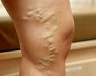
- Compensation for varicose veins. Vessel striae ("stars") appear on the skin. Swelling of the legs at night, cramps, numbness, and pain at night. The healing time of abrasions and abrasions is longer than usual.
- Compensation for varicose veins. The patient continues to worry about pain and increased swelling in the leg. The veins are obviously enlarged and nodules. Skin pigmentation. There are signs of eczema and nutritional disorders.
Pregnant women's pelvic varicose veins also gradually develop. In the first stage, the diameter of the affected blood vessels in any venous plexus of the pelvis does not exceed 5. 0 mm. Second, the uterus or ovaries are involved in this process, and the vascular cavity is 6. 0-10. 0mm. The third feature is that the vein dilates more than 10 mm, causing complete damage to all pelvic venous nerve plexus.
Symptoms of varicose veins
In 80-82% of patients, the disease will feel heavy and tense when it first appears, and the legs will “buzz” and increase at night and during physical exertion. The symptoms of varicose veins gradually increase. As the disease progresses in certain parts of the muscles, pain will appear. This pain first develops with long-term standing and physical labor. In the most severe cases, the pain becomes constant, and the intensity of the pain is so obvious that pregnant women encounter difficulties in independent exercise. Up to 60% of patients noticed calf muscle cramps, up to 40-50%-decreased sensitivity, numbness in the legs, up to 30%-itching.
During the compensatory phase of varicose veins, there are external signs of superficial vein dilation. First, reticular blood vessels and telangiectasia areas ("reticulate" and "stellate") are formed on the skin. Subsequently, the vein pattern became apparent. The veins appear to expand, spiral, and eventually become nodular. The appearance of edema in the ankle and calf area proves the spread of the dilation process to the deep blood vessels. As the varicose veins are decompensated, the skin of the legs looks pigmented, forming eczema. If the pathology appears long before pregnancy and the subcutaneous fat tissue is malnourished, trophic ulcers may occur.
In 4% of patients, the disease affects the veins of the vulva, vagina and small pelvis. For vulvar and vaginal varicose veins, discomfort, swelling, heaviness, and itching are observed in the external genital area. The perineum and labia may swell, and vaginal contact bleeding after intercourse. Pelvic congestion syndrome is manifested as a strain or pain in the lower abdomen, which radiates to the lower back, bones, groin, and external genitals. Dyspareunia is a characteristic of sexual intercourse. In severe cases, dysuria is detected.
Complications
In the absence of proper treatment, the varicose veins of pregnant women will change due to the development of trophic ulcers, erysipelas, thrombophlebitis, superficial and deep vein thrombosis, thromboembolism of the pulmonary artery and other large blood vessels during laborIt's complicated. In 40-45% of cases, placental insufficiency is accompanied by acute and chronic fetal hypoxia. In 25% of patients, abnormal delivery was observed (insufficient labor, dysfunctional myometrial contractions). For vaginal varicose veins, a lot of trauma may occur after delivery. Almost a third of working women have defects in placental separation and placental discharge. The long-term consequences of varicose veins during pregnancy are hemorrhoids, causing chronic venous insufficiency and pelvic pain.
Diagnosis
With the appearance of characteristic skin symptoms, the diagnosis of varicose veins during pregnancy usually does not present any difficulties. The task of the diagnosis stage is to determine the stage and location of vein dilation to rule out other reasons that may cause the stagnation of the lower limb vascular system. The most useful survey method is:
- Chairperson check. This study revealed characteristic changes in the veins of the vulva region and the inner thigh-dilation, tortuosity, nodules. Swelling of the labia and perineum is possible. When viewed in a mirror, the vaginal mucosa looks hypertrophy and hairy. The vaginal vault on palpation with both hands is smooth and usually painful.
- USDG in the venous system. During the ultrasound scan, the shape and diameter of the blood vessel, its length, anatomical location, and wall condition are evaluated. This method allows to determine the branch area, the consistency of the valve device, the patency of the vein, the presence and direction of regurgitation. You can scan the blood vessels of the lower extremities and the inferior vena cava (IVC ultrasound).
- Double scan of blood vessels in the legs. The advantage of a non-invasive method that combines traditional ultrasound and Doppler research is not only to obtain detailed information about blood flow parameters, but also to display the venous network. Double-sided vascular scanning is used to comprehensively assess the state of superficial, perforated and deep blood vessels.
During pregnancy, due to the possibility of negative effects on the fetus, radiological diagnostic methods (varices, selective ovarian imaging, ascending venography of extremities, pelvic venography, CT venography, veins) are only used in a limited rangeScintigraphy, etc. ). In difficult cases, if pelvic varicose is suspected, laparoscopic diagnosis should be performed with caution. The differential diagnosis of varicose veins in the legs includes pregnant women's edema, heart failure, lymphedema, and acute thrombosis of the venous system. Varicose veins of the small pelvis must be distinguished from genital endometriosis, chronic inflammatory pathology of pelvic organs, submucosal and serous uterine fibroids, cysts and other ovarian tumors. In addition to observing obstetricians and gynecologists, it is recommended that patients consult intravenous physicians, cardiologists and oncologists.
Treatment of varicose veins during pregnancy
The main goal of varicose vein treatment for pregnant women is to stop the progression of the disease, reduce the severity of clinical symptoms and prevent possible thromboembolic complications. Non-pharmacological methods are considered advisable, if necessary, supplemented with medication during a safe pregnancy:
- Compression therapy. It is recommended that women who have been diagnosed with varicose veins wear it daily throughout pregnancy, use elastic bandages, special compression tights or 1-2 compression stockings during childbirth and postpartum. Compression therapy by mechanically reducing the diameter of superficial veins can speed up blood flow and reduce swelling and congestion.
- Herbal phlebotonics and phleboprotector. The effect of using this group of drugs is related to increased venous wall tension, decreased permeability, improved microcirculation, blood rheology and lymphatic outflow. The advantage of most bioflavonoids is that they can be used during pregnancy and breastfeeding. Intravenous drugs are available in tablet form or externally.
- Anticoagulants and antiplatelet drugs. When there are signs that there is a tendency to increase coagulation and the threat of developing DIC, drugs with antithrombotic effects should be used with caution. In order to improve the rheology of blood and vascular microcirculation, drugs that can prevent platelet aggregation and have a vascular protective effect have been shown.
It is recommended that pregnant women with varicose veins take special physiotherapy exercises, lymphatic drainage massage, walking according to the dose, and showering in increments every day. Diet correction involves eating foods rich in fiber and vegetable fats. In special cases with severe disease, severe pain syndrome and complications, injection sclerotherapy, microfibrectomy, cross resection, intravascular laser coagulation and other surgical treatments can be used. In most cases, surgical correction is performed at the end of the lactation period.
Delivery strategy
Prediction and prevention
Through timely detection and appropriate treatment, the prognosis is good. As a precaution, it is recommended to have adequate night sleep and regular rest in a supine position throughout the day, with your legs placed on a firm surface at an angle of 30°. Pregnant women with heavy genetics should refuse to wear shoes with heels exceeding 5 cm, limit the time of standing or standing position, and control weight gain.
To prevent varicose veins, take a daily walk and reduce salt intake. It is effective to take vitamin preparations that strengthen the blood vessel wall. According to the indications, patients with varicose veins who plan to become pregnant must undergo surgery to correct the disease.